Introduction
Nontuberculous Mycobacteria (NTM) are diverse group of mycobacteria that are isolated from birds, animals & from environmental sources, such as soil & water. They are opportunistic pathogens occasionally associated with human infection.1
We report a rare case of cutaneous infection caused by Mycobacterium abscessus mimicking actinomycosis.2, 3, 4
Case History
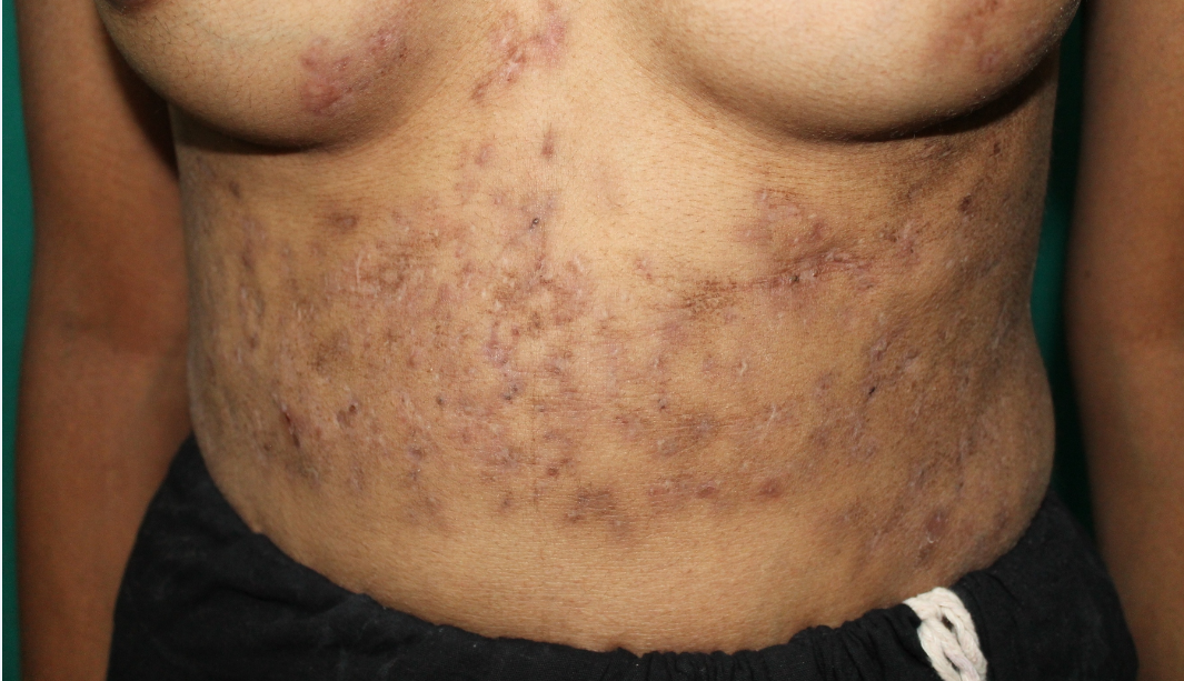
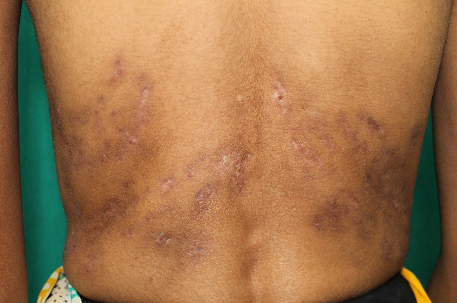
31 years old married female, a known case of rheumatoid arthritis presented with multiple pus-filled lesions, sinuses along with multiple painful firm raised lesions over abdomen, breast and back since past 6 months (Figure 1, Figure 2). On examination it was noted that there were erythematous hyper pigmented tender papules & plaques with atrophic scarring. Induration of sinuses & few pustules were also present. After clinical examination a differential diagnosis of skin TB & actinomycosis was considered. We received skin biopsy specimen for bacteriological, mycobacteriological and fungal workup in laboratory and part of biopsy was also sent to histopathology. On routine culture & sensitivity no organism was isolated.
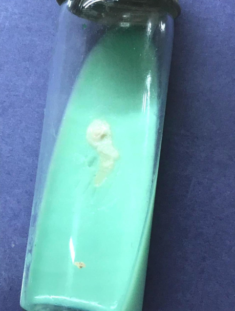
On fifth day, buff colored, moist and no pigmented colonies grown on LJ medium (Figure 3). Gram and ZN stain was performed from the growth. Gram stain showed faintly staining gram positive, short, slender & filamentous bacilli. ZN stain showed acid fast, pleomorphic short thick to filamentous forms (Figure 4). Based on these features it was suspected as a rapid grower atypical mycobacterium. Then the strain on LJ medium was sent to private laboratory for identification of species on MALDI-TOF & antibiotic susceptibility testing. Histopathology revealed suppurative granulomatous tuberculosis inflammation. Acid fast bacilli were present along with Langhans giant cells, macrophages & epithelioid cells. CBNAAT was negative. Based on the histopathology AKT started.
The strain was identified as Mycobacterium abscessus on MALDI-TOF. Then the same strain subjected to Antibiotic susceptibility testing (AST). AST was done by Sensititre broth micro dilution method. Mycobacterium abscessus was sensitive to Cotrimoxazole, Amikacin, Tigecycline, Clarithromycin, Linezolid & Tobramycin. It was intermediate sensitive to Moxifloxacin, Cefoxitin, Doxycycline & Amoxicillin- clavulanic acid. It was found resistant to Ciprofloxacin and Minocycline. It took 6-7 days for identification of species and 15 days for AST. So, after identification and AST patient shifted from AKT to the sensitive drugs. After 10 days of treatment all skin lesions were subsided (Figure 5, Figure 6).
Discussion
According to Runyoun’s classification M. abscessus complex belongs to rapid growing atypical mycobacteria. Other rapid growers are M. Chelonae and M. Fortuitum. M. abscessus was first isolated from a knee abscess in 1952. It had been associated with skin and soft tissue infection, pulmonary infections and hospital acquired infections.5 Cutaneous infections caused by rapid growing mycobacterium typically present with tender, violaceus papules, plaques, nodules or cellulitis on the lower extremities, upper extremities, trunk and rarely on head and neck.6 In our case patient presented with multiple discharging sinuses and erythematous hyper pigmented tender papules and plaques with atrophic scarring over abdomen, breast and back. It is unusual clinical presentation seen with M. abscessus infection. In some case reports similar presentation was seen by M. chelonae infection.7
Rapidly growing mycobacteria on Gram stain appear faintly staining gram positive short and slender bacilli with few filamentous forms. On ZN stain acid fast pleomorphic bacilli ranging from long filamentous forms to short thick rods seen and at times the cells appear beaded or swollen. This group of NTM grows on LJ medium within one week of incubation. Colonies are buff colored in light and dark. The rapid growing mycobacteria are arylsulphatase positive, urease positive, pyrazinamidase positive and grow on 5% NaCl MacConkey’s medium. Newer and rapid methods for identification of all NTM are MALDI-TOF (Matrix Associated Laser Absorption Ionization Time of Flight), DNA probes, Gene sequencing and PCR (Polymerase Chain Reaction).1, 8 In our case we used MALDI-TOF for identification of rapid grower. It was identified as Mycobacterium abscessus.
NTM are resistant to most of the first and second line ant tubercular drugs. So differentiation of NTM from M. tuberculosis is essential. Among all NTM M. abscessus subspecies abscessus is more resistant to antimicrobial agents so species identification is important.1 M. abscessus is an emerging pathogen and cannot be treated with typical antimicrobials like macrolides, fluoroquinolones, aminoglycosides, cefoxitin, imipenem, sulfamethoxazole and tetracyclines (other than tigecycline) without performing AST.8, 9 M.abscessus isolated from this patient was sensitive to Cotrimoxazole, Amikacin, Tigecycline, Clarithromycin, Linezolid & Tobramycin. It was intermediate sensitive to Moxifloxacin, Cefoxitin, Doxycycline & Amoxicillin- clavulanic acid. It was found resistant to Ciprofloxacin and Minocycline. So patient was given clarithromycin, amikacin and cotrimoxazole as per AST report and her lesions were subsided.
Conclusions
NTM should always be considered in a differential diagnosis of skin lesions.
High index of suspicion is required when patient presents with multiple discharging sinuses on skin for diagnosis of NTM cutaneous infections.
Syndromic approach in diagnostic laboratory for any infection is the best approach for correct and early diagnosis of etiological agent.
All parameters like microscopy, culture and histopathology should be considered for diagnosis of mycobacterial infections.
For all acid fast bacilli on microscopy, it is essential to differentiate M. tuberculosis from NTM to start the appropriate treatment.
Among all NTM it is mandatory to identify up to species level and perform AST, as M. abscessus is resistant to typical antimicrobials.


